How To Draw Anatomy Of Heart
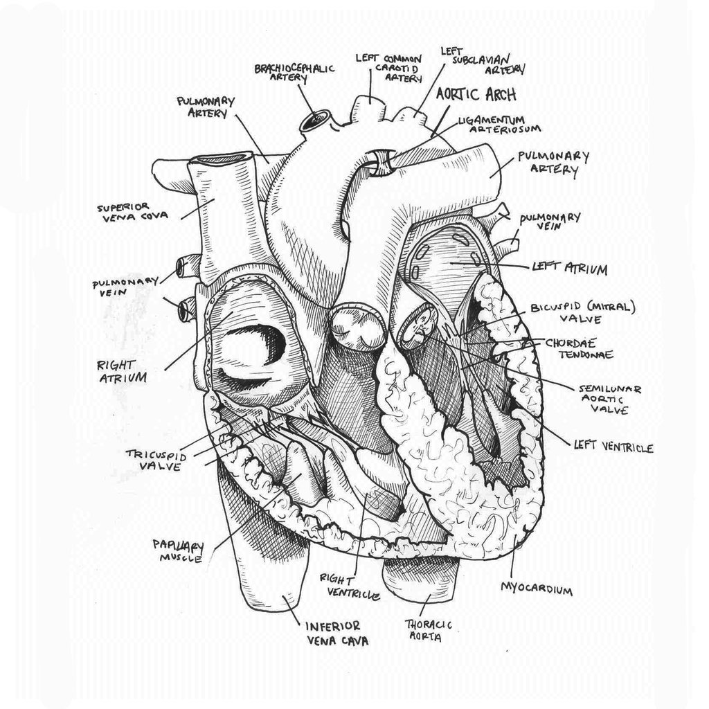
How To Draw Anatomy Of Heart - In this video tutorial you can learn to make perfect. The deoxygenated blood flows through the veins. Understanding its basic anatomy is crucial to understanding how it functions. 48k views 1 year ago cardiovascular. Web the heart is located in the thoracic cavity medial to the lungs and posterior to the sternum. The inferior tip of the heart, known as the apex, rests just superior to the diaphragm. Get free printable coloring page of this drawing. Connect the two with two rounded shapes for the sides of the heart. Web this interactive atlas of human heart anatomy is based on medical illustrations and cadaver photography. The oxygenated blood flows away from the heart. Web the heart is located in the thoracic cavity medial to the lungs and posterior to the sternum. The user can show or hide the anatomical labels which provide a useful tool to create illustrations perfectly adapted for teaching. 271k views 8 years ago. 48k views 1 year ago cardiovascular. The heart is an amazing organ. Web your heart is located in the front of your chest. Start by drawing a rough outline of the heart shape. What does the heart look like and how does it work? The oxygenated blood flows away from the heart. It consists of four main chambers: Dr matt & dr mike. It consists of four main chambers: Web this interactive atlas of human heart anatomy is based on medical illustrations and cadaver photography. Web function and anatomy of the heart made easy using labeled diagrams of cardiac structures and blood flow through the atria, ventricles, valves, aorta, pulmonary arteries veins, superior inferior vena cava, and chambers.. A fun art challenge for everyone. Then, fill in the base of the heart with the right and left ventricles and the right and left atriums. Connect the two with two rounded shapes for the sides of the heart. 3.how to draw a heart diagram 4. Your ribcage protects your heart, everyone’s heart is a slightly different. To begin, draw a triangle shape for the top of the heart. What does the heart look like and how does it work? Method to learn to draw a realistic heart step by step with a pencil. Dr matt & dr mike. It sits slightly behind and to the left of your sternum (breastbone). It sits slightly behind and to the left of your sternum (breastbone). Get free printable coloring page of this drawing. What does the heart look like and how does it work? Dr matt & dr mike. Then, fill in the base of the heart with the right and left ventricles and the right and left atriums. The deoxygenated blood flows through the veins. We will then proceed to shape the heart, slowly refining it with our pencils into a. What does the heart look like and how does it work? Plus, you may just learn something new along the way. Web in this tutorial, we will guide you through the process of drawing the structure of. Web to draw the internal structure of the heart, start by sketching the 2 pulmonary veins to the lower left of the aorta and the bottom of the inferior vena cava slightly to the right of that. Web your heart sure does work hard, but that doesn’t mean you have to work hard to draw it! On its superior end,. The user can show or hide the anatomical labels which provide a useful tool to create illustrations perfectly adapted for teaching. Then, fill in the base of the heart with the right and left ventricles and the right and left atriums. Web the intricate anatomy of the heart can be challenging to grasp, and so i hope you find this. It’s an example of a drawing that can give you a basic idea of what a human heart sketch should be look like. The blood vessels carry the blood through the body. 48k views 1 year ago cardiovascular. The user can show or hide the anatomical labels which provide a useful tool to create illustrations perfectly adapted for teaching. 271k. Plus, you may just learn something new along the way. It sits slightly behind and to the left of your sternum (breastbone). Enhance your artistic skills and create stunning heart illustrations that capture the intricate details. Then, fill in the base of the heart with the right and left ventricles and the right and left atriums. Web this interactive atlas. Sketch out a basic outline of the heart, using our tutorial as a guide. Two atria and two ventricles. Enhance your artistic skills and create stunning heart illustrations that capture the intricate details. The user can show or hide the anatomical labels which provide a useful tool to create illustrations perfectly adapted for teaching. The blood vessels carry the blood. Enhance your artistic skills and create stunning heart illustrations that capture the intricate details. Web this interactive atlas of human heart anatomy is based on medical illustrations and cadaver photography. Web best way to draw and label the heart! Web the main cardiac structures making up the anatomy of the heart include the atria, ventricles, tricuspid valve, mitral valve, aortic valve, and pulmonary valve. Dr matt & dr mike. Without anyone organ not working we would. Understanding its basic anatomy is crucial to understanding how it functions. Web your heart sure does work hard, but that doesn’t mean you have to work hard to draw it! 271k views 8 years ago. It’s an example of a drawing that can give you a basic idea of what a human heart sketch should be look like. Web function and anatomy of the heart made easy using labeled diagrams of cardiac structures and blood flow through the atria, ventricles, valves, aorta, pulmonary arteries veins, superior inferior vena cava, and chambers. There is also a murmurs tab that includes some of the more common murmurs we come across. Next, draw a small curve for the bottom tip of the heart. 48k views 1 year ago cardiovascular. It consists of four main chambers: Add the pulmonary arteries to the triangle shape, one on each side.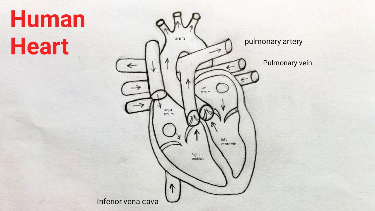
How to draw Human Heart step by step Human Heart diagram easily

How to Draw a Human Heart 11 Steps (with Pictures) wikiHow

How To Draw Human Heart Diagram

Human Heart Drawing & Labelling YouTube

How To Draw Human Heart Diagram

How to Draw the Internal Structure of the Heart (with Pictures)

anatomically correct human heart by NIku Arbabi embroidery
How To Draw An Anatomical Heart The ultimate guide drawboy2

DRAW IT NEAT How to draw human heart labeled Biology Drawing, Study

Sketch of human heart anatomy ,line and color on a checkered background
Connect The Two With Two Rounded Shapes For The Sides Of The Heart.
Then, Fill In The Base Of The Heart With The Right And Left Ventricles And The Right And Left Atriums.
Web The Intricate Anatomy Of The Heart Can Be Challenging To Grasp, And So I Hope You Find This Tool To Be Helpful In Visualizing The Cardiac System.
Your Ribcage Protects Your Heart, Everyone’s Heart Is A Slightly Different.
Related Post: